Dr Tom Burgoyne

Group Leader - UCL Institute of Ophthalmology
Senior Scientist - Royal Brompton Hospital

I have a background in biochemistry, retinal and respiratory biology. My main research interests are in photoreceptor development, health and disc renewal at UCL Institute of Ophthalmology and the development of new diagnostic protocols to diagnose Primary Cilia Dyskinesia (PCD) at the Royal Brompton Hospital. I am currently working on the arrangement of mitochondria and the uptake and trafficking of docosahexaenoic acid (DHA) in photoreceptors, as well as co-supervising a project focussed on the development of a machine learning system to detect ciliary defects in PCD.

Research

My research involves the study of photoreceptors and respiratory cilia by electron microscopy as well as developing image analysis tools and machine learning methods. Due to the SARS-CoV-2 pandemic, we have done some work examining viral infection of airway cells.

Photoreceptors

Photoreceptors are the cells that detect incoming light and enable vision. I have a particular interest in the health and development of these cells. This incluses the structure and renewal of outer segment discs (region of the photoreceptor that detects light) as well as the arrangement of mitochondria and their contact sites with the plasma membrane.

Primary Cilia Dyskinesia

Motile cilia line the airway and move in a beating motion pushing mucus that can contain pathogens away from the lungs where it can be expelled protecting the airways from infection. PCD is a disorder of these cilia leading to increased risk of recurring respiratory tract infections. My work focuses on characterising PCD defects that can be caused by mutations in more than 50 known genes and improving methods to diagnose the disorder.

Coronavirus

Working at the Royal Brompton Hospital, where we specialise in respiratory disease, has allowed me to do SARS-CoV-2 research. This includes studying both infected patient nasal biopsies aand cultured primary airway cells in the hope to better understand how the virus infects cells and replicates.

Caridac Muscle

During my PhD I studied the structure of cardiac muscle. This included comparing it to skeletal muscle and determining the lattice arrangement of muscle filaments using the technique electron tomography.

Electron Microscopy

Much of my work involves electron microscopy, which has allowed me to become an expert in the field. This includes a range of imaging and sample preparation techiques including electron tomography, serial block face scanning electron microscopy, immuno-electron microscopy, high pressure freezing and freeze substitution.

Image Analysis

To assist in the analysis of image data and to enhance the diagnosis of Primary Ciliary Dyskinesia, I have developed a range of programs. Furthermore, at the Royal Brompton Hosptial we are working with Intel to develop a machine learning platform to accurately diagnose PCD.

Publications

Images

The following are a selection of images that relate to some of my published work and research interests.

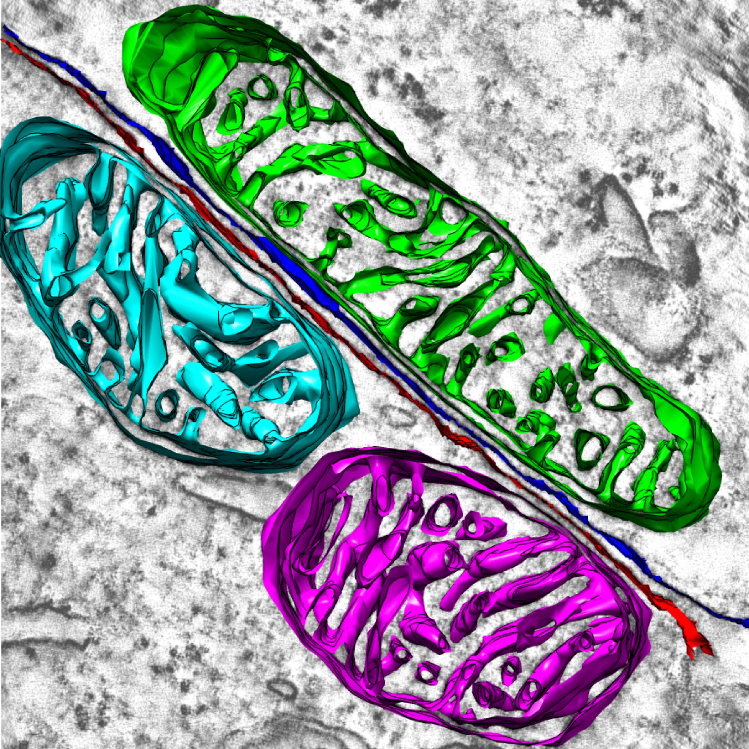
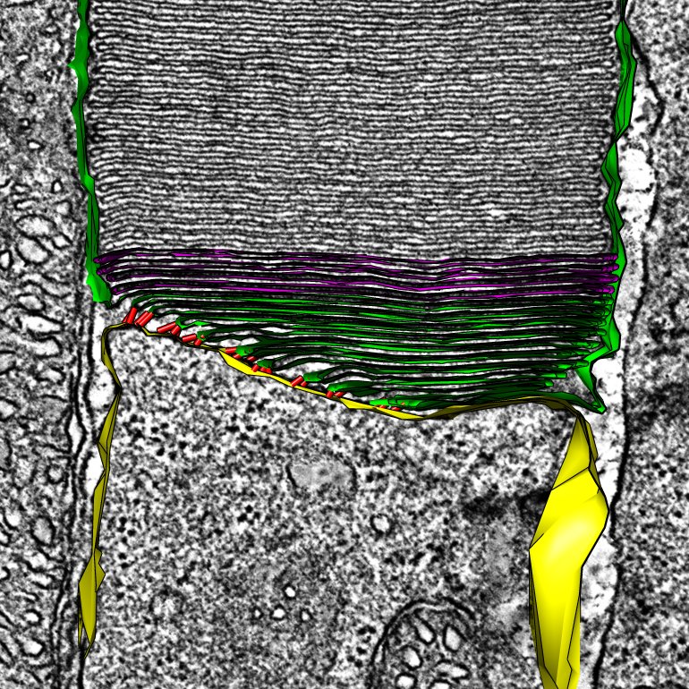
Rod Photoreceptor Disc Renewal

A slice from an electron tomogram that includes an overlay of a model showing the structure of the discs in the light detecting outer segment region of a rod photoreceptor. These disc structures are replaced on a daily basis from evaginations of the plasma membrane (shown in green).

Respiratory Cilia

Electron microscopy cross-sectional image of a human respiratory cilium. These cilia are hair like projections that eminate from the surface of respiratory epithelial cells. They are a type of motile cilia that beat and push mucus that can contain debris and pathogens up the airway where it can be expelled, protecting the airway and lungs.

Vertebrate Muscle Z-Band

Muscle is formed from repeatng units known as sarcomeres. At each end of the sarcomere is a dense structure known as the Z-band. This image shows the structure of small-Square lattice Z-Band in cardiac muscle generated from electron tomography data.

Specialised Arrangement of Mitochondria in Photoreceptor

Electron tomography slice showing two photoreceptor inner segments side-by-side. The plasma membranes of the two cells are shown in red and blue and the mitochondria in green, cyan and purple. Strikely the mitchondria are in contact with the plasma membrane and they are alignment inlcuding their cristae to michondria of neighbouring photoreceptors.

Immuno-gold Labelling of OA1

Immuno-electron microscopy gold labelling of the protein, OA1, expressed in HeLa cells. OA1 localises to multivesicular bodies as shown in this image and plays an important role in the biogenesis of melanosomes. Melanosomes are the organelles that give pigmentation to the skin, but also play a crucial role in preventing cellular damage within the eye caused by light exposure.

SARS-CoV-2 virions

Severe acute respiratory syndrome coronavirus 2 (SARS-CoV-2) virions at the surface of a human airway epithelial cell. Cells were cultured from a nasal brushing biopsy and incubated with virus for 72 hours.

Software

I developed the following programs to perform data and image analysis, some of which are designed to assist in the diagnosis of primary ciliary dyskinesia (PCD).

  • Cilia Counter - Assists in the quantification of ciliary defects to diagnose primary cilia dyskinesia
  • PCD Detect - Toolkit to enhance structural detail through image averaging and classification
  • Sequence Alignment - Simple programs to align nucleotide and protein sequences
  • Filename randomiser - Program that randomises filenames for blinded studies
  • Gold Particle Analyser - Helps analyse gold probes in electron microscopy images
  • Funding

    The following are grants and funding that I have recieved.

    I am extremely grateful for the support that I have had over the years, from funders to those in the lab that have helped me develop as a scientist. A special thanks goes to Moorfields Eye Charity and Royal Brompton and Harefield Hospitals Charity that have provided me funding for preliminary research and publication costs, allowing me to develop my own independent research projects at the UCL Institute of Ophthalmology and Royal Brompton Hospital.